
Automated screening for oxidative or methylation-induced DNA damage in human cells
Article Sidebar
Main Article Content
Abstract
The assessment of genotoxicity upon exposure to chemical and environmental agents plays an important role in basic research as well as in pharmaceutical, chemical, cosmetic and food industry. Low sensitivity and lack of inter-laboratory comparability are considered problematic issues in genotoxicity testing. Moreover, commonly used mutagenicity assays lack information about early and specific genotoxic events.
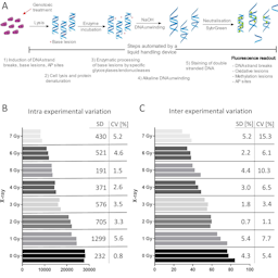
Previously, we developed an automated version of the “Fluorimetric detection of Alkaline DNA Unwinding” (FADU) assay as a high-throughput screening method for the detection of DNA strand breaks in living cells. Here, we report an enzyme-modified version of the cell-based FADU assay (emFADU) for the determination of oxidative and methylation lesions in cellular DNA. Our method is based on the use of formamidopyrimidine DNA glycosylase or human alkyladenine DNA glycosylase for the detection of chemically-induced nucleobase modifications in lysates of immortalized cell lines, growing in suspension or as adherent cells, and in peripheral blood mononuclear cells. We could show that upon treatment with sub-cytotoxic doses of known genotoxins, oxidative and methylation lesions are readily detectable.
This fast, inexpensive, and convenient method could be useful as a high-content screening approach for the sensitive and specific assessment of genotoxicity in human cells. Thus, when implemented in the early compound development in an industrial setting, the emFADU assay could help reduce the number of animals used for toxicity testing. Furthermore, as we established the method for different cell types, this new assay may provide an opportunity for population studies and/or mechanistic research into DNA repair pathways.
Article Details

This work is licensed under a Creative Commons Attribution 4.0 International License.
Articles are distributed under the terms of the Creative Commons Attribution 4.0 International license (http://creativecommons.org/licenses/by/4.0/), which permits unrestricted use, distribution and reproduction in any medium, provided the original work is appropriately cited (CC-BY). Copyright on any article in ALTEX is retained by the author(s).
Abner, C. W., Lau, A. Y., Ellenberger et al. (2001). Base excision and DNA binding activities of human alkyladenine DNA glycosylase are sensitive to the base paired with a lesion. J Biol Chem 276, 13379-13387. doi:10.1074/jbc.M010641200
Aljundi, I. H. (2011). Bromate formation during ozonation of drinking water: A response surface methodology study. Desalination 277, 24-28. doi:10.1016/j.desal.2011.03.090
Beken, S., Kasper, P. and van der Laan, J.W. (2016). Regulatory acceptance of alternative methods in the development and approval of pharmaceuticals. Adv Exp Med Biol 856, 33-64. doi:10.1007/978-3-319-33826-2_3
Beranek, D. T. (1990). Distribution of methyl and ethyl adducts following alkylation with monofunctional alkylating agents. Mutat Res 231, 11-30. doi:10.1016/0027-5107(90)90173-2
Bernstein, C., Bernstein, H., Payne, C. M. et al. (2002). DNA repair/pro-apoptotic dual-role proteins in five major DNA repair pathways: Fail-safe protection against carcinogenesis. Mutat Res 511, 145-178. doi:10.1016/S1383-5742(02)00009-1
Birnboim, H. C. and Jevcak, J. J. (1981). Fluorometric method for rapid detection of DNA strand breaks in human white blood cells produced by low doses of radiation. Cancer Res 41, 1889-1892.
Bouvard, V., Loomis, D., Guyton, K. Z. et al. (2015). Carcinogenicity of consumption of red and processed meat. Lancet Oncol 16, 1599-1600. doi:10.1016/S1470-2045(15)00444-1
Brunborg, G., Rolstadaas, L. and Gutzkow, K. B. (2018). Electrophoresis in the Comet Assay. In O.-M. Boldura and C. Baltă (eds.), Electrophoresis – Life Sciences Practical Applications. InTech. doi:10.5772/intechopen.76880
Cassano, J. C., Roesslein, M., Kaufmann, R. et al. (2020). A novel approach to increase robustness, precision and high-throughput capacity of single cell gel electrophoresis. ALTEX 37, 95-109. doi:10.14573/altex.1906252
Ciccia, A. and Elledge, S. J. (2010). The DNA damage response: Making it safe to play with knives. Mol Cell 40, 179-204. doi:10.1016/j.molcel.2010.09.019
Collins, A. R. (2014). Measuring oxidative damage to DNA and its repair with the comet assay. Biochim Biophys Acta 1840, 794-800. doi:10.1016/j.bbagen.2013.04.022
Cross, A. J. and Sinha, R. (2004). Meat-related mutagens/carcinogens in the etiology of colorectal cancer. Environ Mol Mutagen 44, 44-55. doi:10.1002/em.20030
Enciso, J. M., Gutzkow, K. B., Brunborg, G. et al. (2018). Standardisation of the in vitro comet assay: Influence of lysis time and lysis solution composition on the detection of DNA damage induced by X-rays. Mutagenesis 33, 25-30. doi:10.1093/mutage/gex039
Ersson, C., Møller, P., Forchhammer, L. et al. (2013). An ECVAG inter-laboratory validation study of the comet assay: Inter-laboratory and intra-laboratory variations of DNA strand breaks and FPG-sensitive sites in human mononuclear cells. Mutagenesis 28, 279-286. doi:10.1093/mutage/get001
Hartwig, A., Dally, H. and Schlepegrell, R. (1996). Sensitive analysis of oxidative DNA damage in mammalian cells: Use of the bacterial Fpg protein in combination with alkaline unwinding. Toxicol Lett 88, 85-90. doi:10.1016/0378-4274(96)03722-8
Hoeijmakers, J. H. J. (2009). DNA damage, aging, and cancer. N Engl J Med 361, 1475-1485. doi:10.1056/NEJMra0804615
Hu, C.-W., Chen, C.-M., Ho, H. H. et al. (2012). Simultaneous quantification of methylated purines in DNA by isotope dilution LC-MS/MS coupled with automated solid-phase extraction. Anal Bioanal Chem 402, 1199-1208. doi:10.1007/s00216-011-5559-1
Knudsen, L. E., Smith, A., Törnqvist, E. et al. (2019). Nordic symposium on “toxicology and pharmacology without animal experiments – Will it be possible in the next 10 years?” Basic Clin Pharmacol Toxicol 124, 560-567. doi:10.1111/bcpt.13193
Kumar, A., Dobrovolsky, V., Dhawan A. et al. (2018). Mutagenicity: Assays and Applications. Elsevier. ISBN: 9780128092521
Levin, J. D. and Demple, B. (1990). Analysis of class II (hydrolytic) and class I (beta-lyase) apurinic/apyrimidinic endonucleases with a synthetic DNA substrate. Nucleic Acids Res 18, 5069-5075. doi:10.1093/nar/18.17.5069
Lindahl, T. (1993). Instability and decay of the primary structure of DNA. Nature 362, 709-715. doi:10.1038/362709a0
Moreno-Villanueva, M., Pfeiffer, R., Sindlinger, T. et al. (2009). A modified and automated version of the ‘fluorimetric detection of alkaline DNA unwinding’ method to quantify formation and repair of DNA strand breaks. BMC Biotechnol 9, 39. doi:10.1186/1472-6750-9-39
Moreno-Villanueva, M., Eltze, T., Dressler, D. et al. (2011). The automated FADU-assay, a potential high-throughput in vitro method for early screening of clastogenicity. ALTEX 28, 295-303. doi:10.14573/altex.2011.4.295
Müller, N., Moreno-Villanueva, M., Fischbach, A. et al. (2013). An automated Fpg-based FADU method for the detection of oxidative DNA lesions and screening of antioxidants. Toxicology 310, 15-21. doi:10.1016/j.tox.2013.05.006
Murata, M., Bansho, Y., Inoue, S. et al. (2001). Requirement of glutathione and cysteine in guanine-specific oxidation of DNA by carcinogenic potassium bromate. Chem Res Toxicol 14, 678-685. doi:10.1021/tx000209q
O’Connor, T. R. and Laval, J. (1989). Physical association of the 2,6-diamino-4-hydroxy-5N-formamidopyrimidine-DNA glycosylase of Escherichia coli and an activity nicking DNA at apurinic/apyrimidinic sites. Proc Natl Acad Sci U S A 86, 5222-5226. doi:10.1073/pnas.86.14.5222
Pflaum, M., Will, O. and Epe, B. (1997). Determination of steady-state levels of oxidative DNA base modifications in mammalian cells by means of repair endonucleases. Carcinogenesis 18, 2225-2231. doi:10.1093/carcin/18.11.2225
Sidorenko, V. S., Grollman, A. P., Jaruga, P. et al. (2009). Substrate specificity and excision kinetics of natural polymorphic variants and phosphomimetic mutants of human 8-oxoguanine-DNA glycosylase. FEBS J 276, 5149-5162. doi:10.1111/j.1742-4658.2009.07212.x
Speit, G., Schütz, P., Bonzheim, I. et al. (2004). Sensitivity of the FPG protein towards alkylation damage in the comet assay. Toxicol Lett 146, 151-158. doi:10.1016/j.toxlet.2003.09.010
Sykora, P., Witt, K., Revanna, R. et al. (2018). Next generation high throughput DNA damage detection platform for genotoxic compound screening. Sci Rep 8, 2771. doi:10.1038/s41598-018-20995-w
Tchou, J., Kasai, H., Shibutani, S. et al. (1991). 8-oxoguanine (8-hydroxyguanine) DNA glycosylase and its substrate specificity. Proc Natl Acad Sci U S A 88, 4690-4694. doi:10.1073/pnas.88.11.4690
Wu, Q.-Y., Zhou, Y.-T., Li, W. et al. (2019). Underestimated risk from ozonation of wastewater containing bromide: Both organic byproducts and bromate contributed to the toxicity increase. Water Res 162, 43-52. doi:10.1016/j.watres.2019.06.054
Wyatt, M. D. and Pittman, D. L. (2006). Methylating agents and DNA repair responses: Methylated bases and sources of strand breaks. Chem Res Toxicol 19, 1580-1594. doi:10.1021/tx060164e