
Examination of microcystin neurotoxicity using central and peripheral human neurons
Article Sidebar
Main Article Content
Abstract
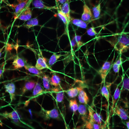
Microcystins (MC) are a group of cyanobacterial toxins that comprises MC-LF and other cyclic heptapeptides, best known as potent hepatotoxicants. Cell culture and epidemiological studies suggest that MC might also affect the nervous system when there is systemic exposure, e.g., via drinking water or food. We asked whether in vitro studies with human neurons could provide estimates on the neurotoxicity hazard of MC-LF. First, we used LUHMES neurons, a well-established test system for neurotoxicants and neuropathological processes. These central nervous system cells express OATP1A2, a presumed carrier of MC-LF, and we observed selective neurite toxicity in the μM range (EC20 = 3.3 μM ≈ 3.3 μg/mL). Transcriptome changes pointed towards attenuated cell maintenance and biosynthetic processes. Prolonged exposure for up to four days did not increase toxicity. As a second model, we used human dorsal root ganglia-like neurons. These peripheral nervous system cells represent parts of the nervous system not protected by the blood-brain barrier in humans. Toxicity was observed in a similar concentration range (EC20 = 7.4 μM). We conclude that MC-LF poses a potential neurotoxic hazard in humans. The adverse effect concentrations observed here were orders of magnitude higher than those presumed to be encountered after normal nutritional or environmental exposure. However, the low μM concentrations found to be toxic are close to levels that may be reached after very excessive algae supplement intake.
Article Details

This work is licensed under a Creative Commons Attribution 4.0 International License.
Articles are distributed under the terms of the Creative Commons Attribution 4.0 International license (http://creativecommons.org/licenses/by/4.0/), which permits unrestricted use, distribution and reproduction in any medium, provided the original work is appropriately cited (CC-BY). Copyright on any article in ALTEX is retained by the author(s).
Altaner, S., Puddick, J., Fessard, V. et al. (2019). Simultaneous detection of 14 microcystin congeners from tissue samples using UPLC-ESI-MS/MS and two different deuterated synthetic microcystins as internal standards. Toxins 11, 388. doi:10.3390/toxins11070388
Altaner, S., Jaeger, S., Fotler, R. et al. (2020). Machine learning prediction of cyanobacterial toxin (microcystin) toxicodynamics in humans. ALTEX 37, 24-36. doi:10.14573/altex.1904031
Azevedo, S. M. F. O., Carmichael, W. W., Jochimsen, E. M. et al. (2002). Human intoxication by microcystins during renal dialysis treatment in caruaru-brazil. Toxicology 181-182, 441-446. doi:10.1016/s0300-483x(02)00491-2
Botes, D. P., Tuinman, A. A., Wessels, P. L. et al. (1984). The structure of cyanoginosin-LA, a cyclic heptapeptide toxin from the cyanobacterium Microcystis aeruginos. J Chem Soc Perkin Trans 1, 2311-2318.
Bronger, H., König, J., Kopplow, K. et al. (2005). ABCC drug efflux pumps and organic anion uptake transporters in human gliomas and the blood-tumor barrier. Cancer Res 65, 11419-11428. doi:10.1158/0008-5472.CAN-05-1271
Buratti, F. M., Manganelli, M., Vichi, S. et al. (2017). Cyanotoxins: Producing organisms, occurrence, toxicity, mechanism of action and human health toxicological risk evaluation. Arch Toxicol 91, 1049-1130. doi:10.1007/s00204-016-1913-6
Carmichael, W. W. (1992). Cyanobacteria secondary metabolites – The cyanotoxins. J Appl Bacteriol 72, 445-459. doi:10.1111/j.1365-2672.1992.tb01858.x
Chen, J., Xie, P., Li, L. et al. (2009). First identification of the hepatotoxic microcystins in the serum of a chronically exposed human population together with indication of hepatocellular damage. Toxicol Sci 108, 81-89. doi:10.1093/toxsci/kfp009
Chen, L. and Xie, P. (2016). Mechanisms of microcystin-induced cytotoxicity and apoptosis. Mini Rev Med Chem 16, 1018-1031. doi:10.2174/1389557516666160219130407
Daneshian, M., Botana, L. M., Dechraoui Bottein, M.-Y. et al. (2013). A roadmap for hazard monitoring and risk assessment of marine biotoxins on the basis of chemical and biological test systems. ALTEX 30, 487-545. doi:10.14573/altex.2013.4.487
Delp, J., Gutbier, S., Klima, S. et al. (2018). A high-throughput approach to identify specific neurotoxicants/ developmental toxicants in human neuronal cell function assays. ALTEX 35, 235-253. doi:10.14573/altex.1712182
Delp, J., Funke, M., Rudolf, F. et al. (2019). Development of a neurotoxicity assay that is tuned to detect mitochondrial toxicants. Arch Toxicol 93, 1585-1608. doi:10.1007/s00204-019-02473-y
Desprez, B., Birk, B., Blaauboer, B. et al. (2019). A mode-of-action ontology model for safety evaluation of chemicals: Outcome of a series of workshops on repeated dose toxicity. Toxicol In Vitro 59, 44-50. doi:10.1016/j.tiv.2019.04.005
Devos, D., Moreau, C., Devedjian, J. C. et al. (2014). Targeting chelatable iron as a therapeutic modality in Parkinson’s disease. Antioxid Redox Signal 21, 195-210. doi:10.1089/ars.2013.5593
Dietrich, D. and Hoeger, S. (2005). Guidance values for microcystins in water and cyanobacterial supplement products (blue-green algal supplements): A reasonable or misguided approach? Toxicol Appl Pharmacol 203, 273-289. doi:10.1016/j.taap.2004.09.005
Feurstein, D., Holst, K., Fischer, A. et al. (2009). OATP-associated uptake and toxicity of microcystins in primary murine whole brain cells. Toxicol Appl Pharmacol 234, 247-255. doi:10.1016/j.taap.2008.10.011
Feurstein, D., Stemmer, K., Kleinteich, J. et al. (2011). Microcystin congener- and concentration-dependent induction of murine neuron apoptosis and neurite degeneration. Toxicol Sci 124, 424-431. doi:10.1093/toxsci/kfr243
Fischer, W. J. and Dietrich, D. R. (2000). Pathological and biochemical characterization of microcystin-induced hepatopancreas and kidney damage in carp (Cyprinus carpio). Toxicol Appl Pharmacol 164, 73-81. doi:10.1006/taap.1999.8861
Fischer, W. J., Altheimer, S., Cattori, V. et al. (2005). Organic anion transporting polypeptides expressed in liver and brain mediate uptake of microcystin. Toxicol Appl Pharmacol 203, 257-263. doi:10.1016/j.taap.2004.08.012
Gao, Y.-s., Hubbert, C. C., Lu, J. et al. (2007). Histone deacetylase 6 regulates growth factor-induced actin remodeling and endocytosis. Mol Cell Biol 27, 8637-8647. doi:10.1128/MCB.00393-07
Gutbier, S., Spreng, A.-S., Delp, J. et al. (2018). Prevention of neuronal apoptosis by astrocytes through thiol-mediated stress response modulation and accelerated recovery from proteotoxic stress. Cell Death Differ 25, 2101-2117. doi:10.1038/s41418-018-0229-x
Hagenbuch, B. and Meier, P. J. (2003). The superfamily of organic anion transporting polypeptides. Biochim Biophys Acta 1609, 1-18. doi:10.1016/s0005-2736(02)00633-8
Herrera, N., Herrera, C., Ortíz, I. et al. (2018). Genotoxicity and cytotoxicity of three microcystin-LR containing cyanobacterial samples from Antioquia, Colombia. Toxicon 154, 50-59. doi:10.1016/j.toxicon.2018.09.011
Heussner, A. H., Mazija, L., Fastner, J. et al. (2012). Toxin content and cytotoxicity of algal dietary supplements. Toxicol Appl Pharmacol 265, 263-271. doi:doi:10.1016/j.taap.2012.10.005
Hinojosa, M. G., Gutiérrez-Praena, D., Prieto, A. I. et al. (2019). Neurotoxicity induced by microcystins and cylindrospermopsin: A review. Sci Total Environ 668, 547-565. doi:10.1016/j.scitotenv.2019.02.426
Hoelting, L., Klima, S., Karreman, C. et al. (2016). Stem cell-derived immature human dorsal root ganglia neurons to identify peripheral neurotoxicants. Stem Cells Transl Med 5, 476-487. doi:10.5966/sctm.2015-0108
Höllerhage, M., Moebius, C., Melms, J. et al. (2017). Protective efficacy of phosphodiesterase-1 inhibition against alpha-synuclein toxicity revealed by compound screening in LUHMES cells. Sci Rep 7, 11469-11469. doi:10.1038/s41598-017-11664-5
House, J. S., Grimm, F. A., Jima, D. D. et al. (2017). A pipeline for high-throughput concentration response modeling of gene expression for toxicogenomics. Front Genet 8, 168. doi:10.3389/fgene.2017.00168
Hu, Y., Chen, J., Fan, H. et al. (2016). A review of neurotoxicity of microcystins. Environ Sci Pollut Res Int 23, 7211-7219. doi:10.1007/s11356-016-6073-y
Jochimsen, E. M., Carmichael, W. W., An, J. S. et al. (1998). Liver failure and death after exposure to microcystins at a hemodialysis center in Brazil. N Engl J Med 338, 873-878. doi:10.1056/NEJM199803263381304
Karreman, C., Klima, S., Holzer, A. K. et al. (2020). CaFFEE: A program for evaluating time courses of Ca2+ dependent signal changes of complex cells loaded with fluorescent indicator dyes. ALTEX 37, 332-336. doi:10.14573/altex.2003191
Krug, A. K., Balmer, N. V., Matt, F. et al. (2013). Evaluation of a human neurite growth assay as specific screen for developmental neurotoxicants. Arch Toxicol 87, 2215-2231. doi:10.1007/s00204-013-1072-y
Li, Y., Chen, J.-a., Zhao, Q. et al. (2011). A cross-sectional investigation of chronic exposure to microcystin in relationship to childhood liver damage in the Three Gorges Reservoir Region, China. Environ Health Perspect 119, 1483-1488. doi:10.1289/ehp.1002412
Livak, K. J. and Schmittgen, T. D. (2001). Analysis of relative gene expression data using real-time quantitative PCR and the 2-ΔΔC(T) method. Methods 25, 402-408. doi:10.1006/meth.2001.1262
Lohren, H., Blagojevic, L., Fitkau, R. et al. (2015). Toxicity of organic and inorganic mercury species in differentiated human neurons and human astrocytes. J Trace Elem Med Biol 32, 200-208. doi:10.1016/j.jtemb.2015.06.008
Lotharius, J., Falsig, J., van Beek, J. et al. (2005). Progressive degeneration of human mesencephalic neuron-derived cells triggered by dopamine-dependent oxidative stress is dependent on the mixed-lineage kinase pathway. J Neurosci 25, 6329-6342. doi:10.1523/JNEUROSCI.1746-05.2005
Love, M. I., Huber, W. and Anders, S. (2014). Moderated estimation of fold change and dispersion for RNA-seq data with DESeq2. Genome Biol 15, 550. doi:10.1186/s13059-014-0550-8
MacKintosh, C., Beattie, K. A., Klumpp, S. et al. (1990). Cyanobacterial microcystin-LR is a potent and specific inhibitor of protein phosphatases 1 and 2A from both mammals and higher plants. FEBS Lett 264, 187-192. doi:10.1016/0014-5793(90)80245-e
Matelski, L., Morgan, R. K., Grodzki, A. C. et al. (2020). Effects of cytokines on nuclear factor-kappa B, cell viability, and synaptic connectivity in a human neuronal cell line. Mol Psychiatry. doi:10.1038/s41380-020-0647-2
Metsalu, T. and Vilo, J. (2015). Clustvis: A web tool for visualizing clustering of multivariate data using principal component analysis and heatmap. Nucleic Acids Res 43, W566-W570. doi:10.1093/nar/gkv468
Nishiwaki-Matsushima, R., Ohta, T., Nishiwaki, S. et al. (1992). Liver tumor promotion by the cyanobacterial cyclic peptide toxin microcystin-LR. J Cancer Res Clin Oncol 118, 420-424. doi:10.1007/bf01629424
Pouria, S., de Andrade, A., Barbosa, J. et al. (1998). Fatal microcystin intoxication in haemodialysis unit in Caruaru, Brazil. Lancet 352, 21-26. doi:10.1016/s0140-6736(97)12285-1
Raudvere, U., Kolberg, L., Kuzmin, I. et al. (2019). g:Profiler: A web server for functional enrichment analysis and conversions of gene lists (2019 update). Nucleic Acids Res 47, W191-W198. doi:10.1093/nar/gkz369
Scholz, D., Pöltl, D., Genewsky, A. et al. (2011). Rapid, complete and large-scale generation of post-mitotic neurons from the human luhmes cell line. J Neurochem 119, 957-971. doi:10.1111/j.1471-4159.2011.07255.x
Scholz, D., Chernyshova, Y., Ückert, A.-K. et al. (2018). Reduced Aβ secretion by human neurons under conditions of strongly increased BACE activity. J Neurochem 147, 256-274. doi:10.1111/jnc.14467
Singh, N., Lawana, V., Luo, J. et al. (2018). Organophosphate pesticide chlorpyrifos impairs STAT1 signaling to induce dopaminergic neurotoxicity: Implications for mitochondria mediated oxidative stress signaling events. Neurobiol Dis 117, 82-113. doi:10.1016/j.nbd.2018.05.019
Skirzewski, M., Karavanova, I., Shamir, A. et al. (2018). ErbB4 signaling in dopaminergic axonal projections increases extracellular dopamine levels and regulates spatial/working memory behaviors. Mol Psychiatry 23, 2227-2237. doi:10.1038/mp.2017.132
Spoof, L. and Catherine, A. (2016). Appendix 3: Tables of microcystins and nodularins. In J. Meriluoto, L. Spoof and G. A. Codd (eds.), Handbook of Cyanobacterial Monitoring and Cyanotoxin Analysis (526-537). John Wiley & Sons, Ltd. doi:10.1002/9781119068761.app3
Stiegler, N. V., Krug, A. K., Matt, F. et al. (2011). Assessment of chemical-induced impairment of human neurite outgrowth by multiparametric live cell imaging in high-density cultures. Toxicol Sci 121, 73-87. doi:10.1093/toxsci/kfr034
Svirčev, Z., Lalić, D., Bojadžija Savić, G. et al. (2019). Global geographical and historical overview of cyanotoxin distribution and cyanobacterial poisonings. Arch Toxicol 93, 2429-2481. doi:10.1007/s00204-019-02524-4
Tong, Z.-B., Huang, R., Wang, Y. et al. (2018). The Toxmatrix: Chemo-genomic profiling identifies interactions that reveal mechanisms of toxicity. Chem Res Toxicol 31, 127-136. doi:10.1021/acs.chemrestox.7b00290
Valério, E., Vasconcelos, V. and Campos, A. (2016). New insights on the mode of action of microcystins in animal cells – A review. Mini Rev Med Chem 16, 1032-1041. doi:10.2174/1389557516666160219130553
Waldmann, T., Rempel, E., Balmer, N. V. et al. (2014). Design principles of concentration-dependent transcriptome deviations in drug-exposed differentiating stem cells. Chem Res Toxicol 27, 408-420. doi:10.1021/tx400402j
Weng, M. K., Zimmer, B., Pöltl, D. et al. (2012). Extensive transcriptional regulation of chromatin modifiers during human neurodevelopment. PLoS One 7, e36708-e36708. doi:10.1371/journal.pone.0036708
Weng, M. K., Natarajan, K., Scholz, D. et al. (2014). Lineage-specific regulation of epigenetic modifier genes in human liver and brain. PLoS One 9, e102035-e102035. doi:10.1371/journal.pone.0102035
Witt, B., Meyer, S., Ebert, F. et al. (2017). Toxicity of two classes of arsenolipids and their water-soluble metabolites in human differentiated neurons. Arch Toxicol 91, 3121-3134. doi:10.1007/s00204-017-1933-x
Yoshizawa, S., Matsushima, R., Watanabe, M. F. et al. (1990). Inhibition of protein phosphatases by microcystins and nodularin associated with hepatotoxicity. J Cancer Res Clin Oncol 116, 609-614. doi:10.1007/bf01637082