
In vitro model of neurotrauma using the chick embryo to test regenerative bioimplantation
Article Sidebar
Main Article Content
Abstract
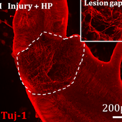
Effective repair of spinal cord injury sites remains a major clinical challenge. One promising strategy is the implantation of multifunctional bioscaffolds to enhance nerve fiber growth, guide regenerating tissue, and modulate scarring/inflammation processes. Given their multifunctional nature, such implants require testing in models which replicate the complex neuropathological responses of spinal injury sites. This is often achieved using live, adult animal models of spinal injury. However, these have substantial drawbacks for developmental testing, including the requirement for large numbers of animals, costly infrastructure, high levels of expertise, and complex ethical processes. As an alternative, we show that organotypic spinal cord slices can be derived from the E14 chick embryo and cultured with high viability for at least 24 days, with major neural cell types detected. A transecting injury could be reproducibly introduced into the slices and characteristic neuropathological responses similar to those in adult spinal cord injury observed at the lesion margin. This included aligned astrocyte morphologies and upregulation of glial fibrillary acidic protein in astrocytes, microglial infiltration into the injury cavity, and limited nerve fiber outgrowth. Bioimplantation of a clinical grade scaffold biomaterial was able to modulate these responses, disrupting the astrocyte barrier, enhancing nerve fiber growth, and supporting immune cell invasion. Chick embryos are inexpensive and simple, requiring facile methods to generate the neurotrauma model. Our data show the chick embryo spinal cord slice system could be a replacement spinal injury model for laboratories developing new tissue engineering solutions.
Plain language summary
Spinal cord injury can be highly debilitating for patients and carers. Repair of the spinal cord is therefore a major clinical goal but is challenging owing to the complex pathology of the injury site. Researchers need to mimic this complexity in the laboratory to test new therapies in realistic environments. Traditionally, researchers use live, adult animal models to achieve this, which are highly traumatic, variable, and costly. Here, we show an alternative system based on slices of spinal cord tissue from chick embryos that could partially replace live adult animal testing. We show slices can be grown in a dish and reproducibly injured, with complex pathology replicated. Further, we modified cell responses in the injury through interface with a novel implant, potentially improving repair. The model is cost-effective, simple, and associated with less animal suffering than live adult animal experiments.
Article Details

This work is licensed under a Creative Commons Attribution 4.0 International License.
Articles are distributed under the terms of the Creative Commons Attribution 4.0 International license (http://creativecommons.org/licenses/by/4.0/), which permits unrestricted use, distribution and reproduction in any medium, provided the original work is appropriately cited (CC-BY). Copyright on any article in ALTEX is retained by the author(s).
Boni, R., Ali, A., Shavandi, A. et al. (2018). Current and novel polymeric biomaterials for neural tissue engineering. J Biomed Sci 25, 90. doi:10.1186/s12929-018-0491-8
Bradbury, E. J. and Burnside, E. R. (2019). Moving beyond the glial scar for spinal cord repair. Nat Commun 10, 3879. doi:10.1038/s41467-019-11707-7
Calderó, J., Brunet, N., Ciutat, D. et al. (2009). Development of microglia in the chick embryo spinal cord: Implications in the regulation of motoneuronal survival and death. J Neurosci Res 87, 2447-2466. doi:10.1002/jnr.22084
Doblado, L. R., Martínez-Ramos, C. and Pradas, M. M. (2021). Biomaterials for neural tissue engineering. Front Nanotechnol 3. doi:10.3389/fnano.2021.643507
Domínguez-Bajo, A., González-Mayorga, A., López-Dolado, E. et al. (2020). Graphene oxide microfibers promote regenerative responses after chronic implantation in the cervical injured spinal cord. ACS Biomater Sci Eng 6, 2401-2414. doi:10.1021/acsbiomaterials.0c00345
Ferretti, P. and Whalley, K. (2008). Successful neural regeneration in amniotes: The developing chick spinal cord. Cell Mol Life Sci 65, 45-53. doi:10.1007/s00018-007-7430-2
Führmann T., Anandakumaran, P. N. and Shoichet, M. S. (2017). Combinatorial therapies after spinal cord injury: How can biomaterials help? Adv Healthc Mater 6, 1601130. doi:10.1002/adhm.201601130
Galli, R., Sitoci-Ficici, K. H., Uckermann, O. et al. (2018). Label-free multiphoton microscopy reveals relevant tissue changes induced by alginate hydrogel implantation in rat spinal cord injury. Sci Rep 8, 10841. doi:10.1038/s41598-018-29140-z
Guijarro-Belmar, A., Viskontas, M., Wei, Y. et al. (2019). Epac2 elevation reverses inhibition by chondroitin sulfate proteoglycans in vitro and transforms postlesion inhibitory environment to promote axonal outgrowth in an ex vivo model of spinal cord injury. J Neurosci 39, 8330-8346. doi:10.1523/jneurosci.0374-19.2019
Hasan, S. J., Keirstead, H. S., Muir, G. D. et al. (1993). Axonal regeneration contributes to repair of injured brainstem-spinal neurons in embryonic chick. J Neurosci 13, 492-507. doi:10.1523/jneurosci.13-02-00492.1993
Himmels, P., Paredes, I., Adler, H. et al. (2017). Motor neurons control blood vessel patterning in the developing spinal cord. Nat Commun 8, 14583. doi:10.1038/ncomms14583
Hurtado, A., Cregg, J. M., Wang, H. B. et al. (2011). Robust CNS regeneration after complete spinal cord transection using aligned poly-L-lactic acid microfibers. Biomaterials 32, 6068-6079. doi:10.1016/j.biomaterials.2011.05.006
Kourgiantaki, A., Tzeranis, D. S., Karali, K. et al. (2020). Neural stem cell delivery via porous collagen scaffolds promotes neuronal differentiation and locomotion recovery in spinal cord injury. NPJ Regen Med 5, 12. doi:10.1038/s41536-020-0097-0
Lee, J. H., Shin, H., Shaker, M. R. et al. (2022). Production of human spinal-cord organoids recapitulating neural-tube morphogenesis. Nat Biomed Eng 6, 435-448. doi:10.1038/s41551-022-00868-4
Mogas Barcons, A., Chari, D. M. and Adams, C. (2021). Enhancing the regenerative potential of stem cell-laden, clinical-grade implants through laminin engineering. Mater Sci Eng C 123, 111931. doi:10.1016/j.msec.2021.111931
Ogaki, A., Ikegaya, Y. and Koyama, R. (2022). Replacement of mouse microglia with human induced pluripotent stem cell (hiPSC)-derived microglia in mouse organotypic slice cultures. Front Cell Neurosci 16, 918442. doi:10.3389/fncel.2022.918442
Sharma, A. L., Wang H., Zhang, Z. et al. (2022). HIV promotes neurocognitive impairment by damaging the hippocampal microvessels. Mol Neurobiol 59, 4966-4986. doi:10.1007/s12035-022-02890-8
Shimizu, I., Oppenheim, R. W., O’Brien, M. et al. (1990). Anatomical and functional recovery following spinal cord transection in the chick embryo. J Neurobiol 21, 918-937. doi:10.1002/neu.480210609
Tubby, K. C., Norval, D. and Price, S. R. (2013). Chicken embryo spinal cord slice culture protocol. J Vis Exp 73, 50295. doi:10.3791/50295
Walsh, C. M., Wychowaniec, J. K., Costello, L. et al. (2023). An in vitro and ex vivo analysis of the potential of GelMA hydrogels as a therapeutic platform for preclinical spinal cord injury. Adv Healthc Mater 12, e2300951. doi:10.1002/adhm.202300951
Weightman, A. P., Pickard, M. R., Yang, Y. et al. (2014). An in vitro spinal cord injury model to screen neuroregenerative materials. Biomaterials 35, 3756-3765. doi:10.1016/j.biomaterials.2014.01.022
Whalley, K., O’Neill, P. and Ferretti, P. (2006). Changes in response to spinal cord injury with development: Vascularization, hemorrhage and apoptosis. Neuroscience 137, 821-832. doi:10.1016/j.neuroscience.2005.07.064
Wu, G. H., Shi, H. J., Che, M. T. et al. (2018). Recovery of paralyzed limb motor function in canine with complete spinal cord injury following implantation of MSC-derived neural network tissue. Biomaterials 181, 15-34. doi:10.1016/j.biomaterials.2018.07.010
Xue, W., Li, B., Liu, H. et al. (2022). Generation of dorsoventral human spinal cord organoids via functionalizing composite scaffold for drug testing. iScience 26, 105898. doi:10.1016/j.isci.2022.105898
Yang, C., Li, X., Li, S. et al. (2019). Organotypic slice culture based on in ovo electroporation for chicken embryonic central nervous system. J Cell Mol Med 23, 1813-1826. doi:10.1111/jcmm.14080
Yang, Z., Zhang, A., Duan, H. et al. (2015). NT3-chitosan elicits robust endogenous neurogenesis to enable functional recovery after spinal cord injury. Proc Natl Acad Sci U S A 112, 13354-13359. doi:10.1073/pnas.1510194112